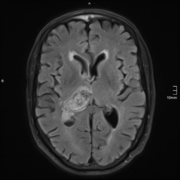
Brain Imaging

Hi Everyone,
Thank you to everyone for joining us at our most recent session!
Exciting news: we are expanding the journal club and looking to recruit new committee members. If you are interested in joining, please complete the Google Form.
Deadline to apply: 30th December.
Please see below a summary of the papers discussed
Imaging in Acute Ischaemic Stroke: Assessing Findings in Light of Evolving Therapies
- Stroke is one of the leading causes of disability and death in the UK, with 85% being ischaemic and 15% haemorrhagic.
- Timely imaging is crucial for management:
- Non-contrast CT (NCCT):
- First-line imaging to exclude haemorrhage.
- Limitations: Poor sensitivity for early ischaemic changes.
- ASPECTS (Alberta Stroke Program Early CT Score) helps assess prognosis, especially for MCA infarcts, with lower scores indicating worse outcomes.
- CT Angiography (CTA): Identifies large vessel occlusions and collateral circulation, guiding suitability for thrombectomy.
- CT Perfusion (CTP): Differentiates salvageable penumbra from the infarct core, key for decisions on thrombectomy up to 24 hours post-stroke.
- Penumbra: Increased mean transit time (MTT), normal/increased cerebral blood volume (CBV).
- Infarct core: Markedly reduced CBV and cerebral blood flow (CBF).
- MRI: Increasingly important for posterior circulation strokes and distinguishing viable penumbra with DWI-FLAIR mismatch.
- Non-contrast CT (NCCT):
- Emerging AI tools may enhance detection and intervention outcomes.
The Role of Neuroimaging in Early Dementia Diagnosis
- Dementia affects 55 million people worldwide, with over 60% from low- to middle-income countries.
- Imaging plays a pivotal role in early diagnosis, helping optimise management and support for patients and families:
- Structural MRI: Preferred over CT due to superior soft tissue contrast and no radiation.
- Identifies characteristic patterns of brain atrophy to differentiate dementia types (e.g., hippocampal atrophy in Alzheimer’s).
- Volumetric analysis improves sensitivity and specificity, particularly in advanced cases.
- Functional MRI: Assesses brain activity:
- AD shows reduced connectivity in the dorsal visual stream.
- FTD displays impaired auditory and visual processing pathways.
- FDG-PET: Measures glucose metabolism, identifying areas of hypometabolism characteristic of different dementias.
- AD: Hypometabolism in temporal/parietal lobes.
- FTD: Frontal lobe hypometabolism.
- DLB: Occipital hypometabolism.
- Amyloid PET: Visualises amyloid plaques in AD, aiding early and differential diagnosis, especially in early-onset cases.
- Structural MRI: Preferred over CT due to superior soft tissue contrast and no radiation.
- Emerging tools like tau PET imaging and blood-brain barrier studies are advancing diagnostic precision.
If you have any questions, feel free to contact the team. We look forward to seeing you at our next session!