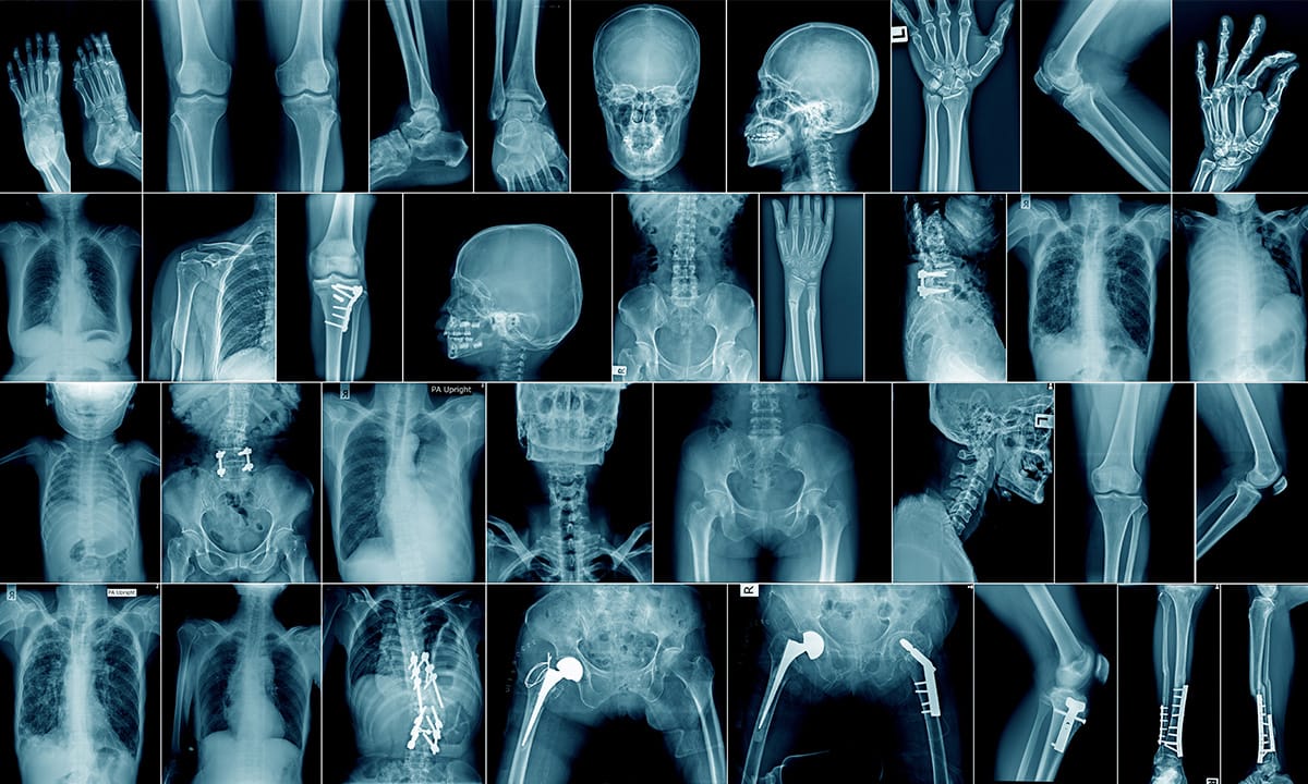
🩻 BRJC Session 5 Recap: Abdominal & Musculoskeletal X-Rays – A Practical Guide
Thank you to everyone who joined Session 5 of the British Radiology Journal Club! This practical and visually rich session was led by Dr. Maryam Ikram (PGR4 Radiology, INMOL Hospital, Pakistan) and focused on the essentials of abdominal and musculoskeletal X-ray interpretation — from foundational anatomy to emergency pathology recognition.
🗓️ Event Overview
📍 Topic: Abdominal & MSK X-Rays – A Practical Guide
🎙️ Speaker: Dr. Maryam Ikram
🕖 Time: 7 PM BST
🧠 Session Highlights
Designed for clinical relevance and practical application, this session offered a systematic approach to both abdominal and MSK radiography, with an emphasis on pattern recognition and common pitfalls.
🔍 Abdominal X-Ray Interpretation
🧩 Anatomy & Normal Patterns
- Gas patterns:
- Small bowel: central with valvulae conniventes (full-width mucosal folds)
- Large bowel: peripheral, with haustra (non-continuous folds)
- Bowel diameter ("3/6/9 Rule"):
- Small bowel ≤ 3 cm
- Colon ≤ 6 cm
- Caecum ≤ 9 cm
- Faecal pattern: mottled gas in colon; absence suggests obstruction
🚨 Key Pathologies
- Small Bowel Obstruction:
- Dilated loops >3cm
- Multiple air-fluid levels
- No rectal gas
- "Coiled-spring" appearance
- Volvulus:
- Sigmoid – coffee-bean sign
- Cecal – kidney-shaped mass, displaced cecum
- Pneumoperitoneum:
- Free air under diaphragm on erect AXR
- Rigler's sign: gas on both sides of bowel wall
- Renal stones vs Phleboliths:
- Renal stones are dense with no lucency
- Phleboliths have central lucency
💀 Musculoskeletal X-Ray Essentials
🔎 Normal Features
- Long bones: cortex, medullary canal
- Joints: assess joint space, subchondral bone
- Soft tissues: look for swelling, fat pads
🦴 Fracture Types
- Transverse, oblique, spiral, comminuted
- Greenstick: common in paediatrics
- Fat pad signs (elbow):
- Anterior sail sign: raised anterior fat pad = suspect fracture
- Posterior fat pad: often indicates occult fracture
🧪 Interactive Case Review
Participants engaged in interpreting real X-ray cases:
- Case 1–2: Small and large bowel obstructions with classic imaging signs
- Case 3: Pneumoperitoneum with free subdiaphragmatic air
- Additional MSK and emergency cases were reviewed interactively, reinforcing a structured image analysis approach
🧠 Key Takeaways
- Always start with normal anatomy
- AXR is a screening tool; CT is diagnostic
- In MSK, know your red flags: hidden fractures, effusions, displaced fat pads
🛠️ Recommended Resources
- Radiopaedia.org
- LearningRadiology.com
- GeekyMedics
- FRCR anatomy apps/modules
💬 Join the BRJC Community
Get access to upcoming events, learning materials, and peer discussion.
👉 Join our WhatsApp group
📌 What’s Next?
Stay tuned for Session 6, with more practical radiology insights and hands-on learning.
📲 Stay Connected
📸 Instagram: @official.brjc
📘 Facebook: British Radiology Journal Club
Thanks again for being part of the BRJC community. See you at the next session!